
Fascia in Horses – Danish veterinary exploring uncharted territory
- In 2015 veterinary Vibeke S Elbrønd published the first report on Fascia and horses.
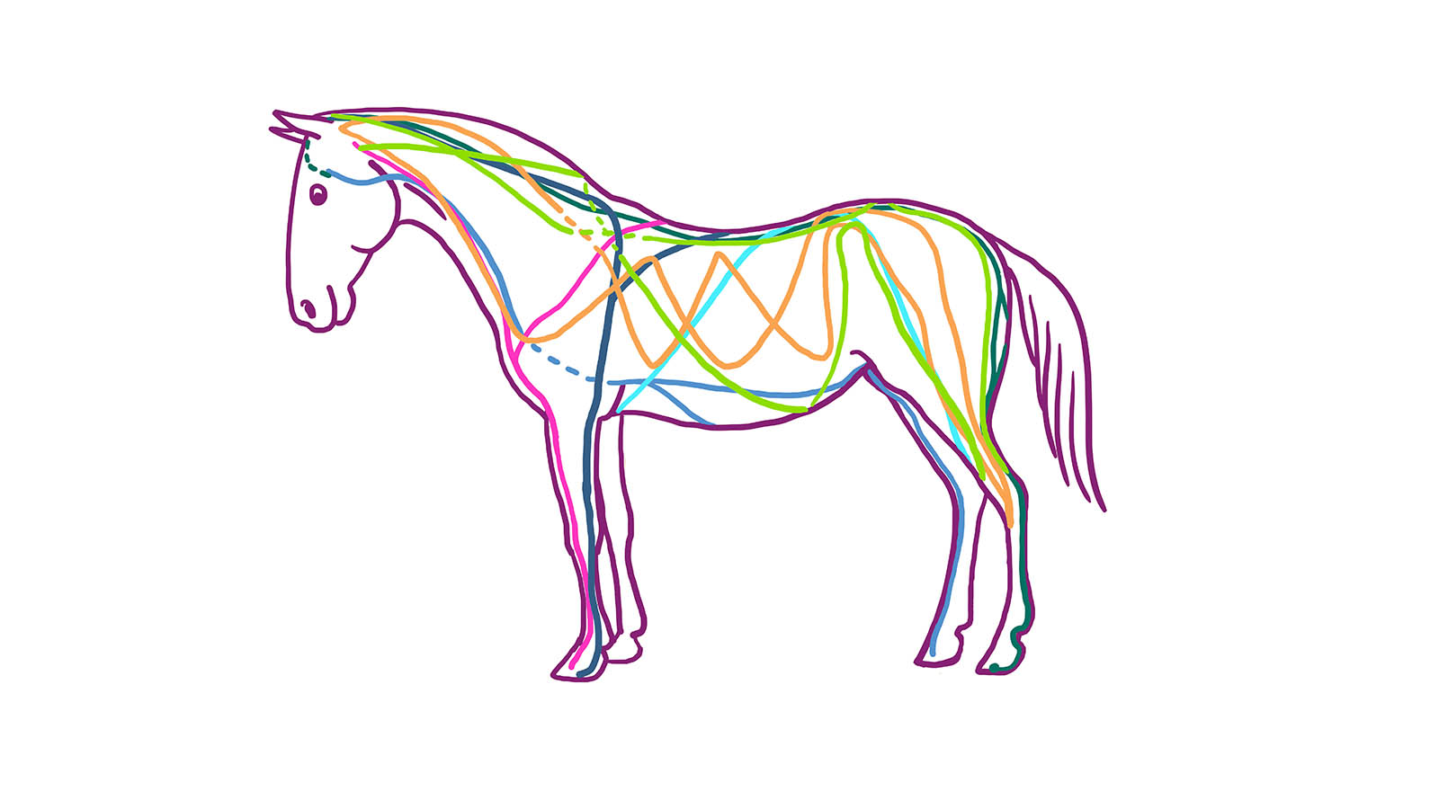
- Through autopsy she found that the horse has the same kind of chains and networks of connective tissue through the body, as found in humans by Tom Myers.
- They are called “Myofascial chains in horses”. Since the horse is a quadruped these lines – also described as chains – are 90% consistent with those in humans.
- In a parallell study she used the Atlasbalans M1 Fascia Treatment machine to prove the existence of the myofascial chains. The study showed how the vibrations spread from the neck to the back and down to the hoofs
- The studies were presented at the Fascia Research Congress in Washington DC, September 2015.
- PDF: Myofascia – the unexplored tissue
- PDF: Multi-Frequency Bioimpedance and Myofascial Release Therapy: An equine Atlasbalans M1 validation study
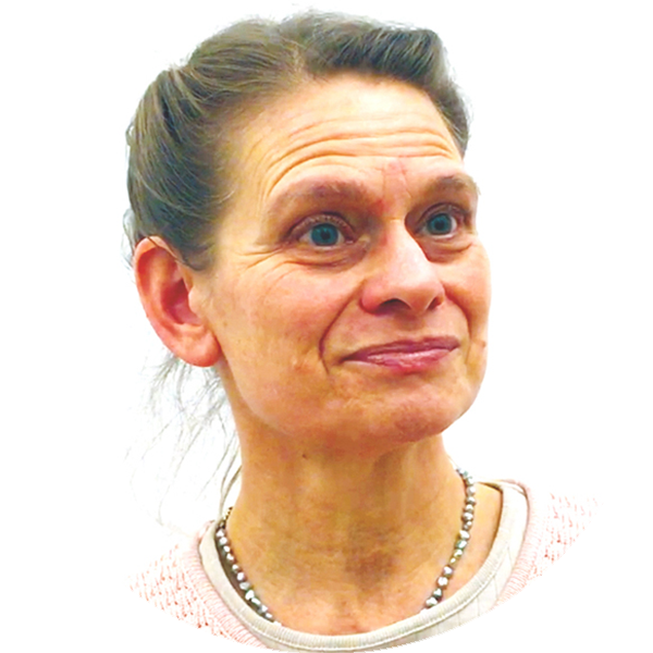
Video: Interview with Dr. Vibeke Sødring Elbrønd
Myofascia – the unexplored tissue
The precise functional role of connective tissue, and especially that of Myofascia, remains largely unexplored. With this in mind, the present study, Myofascia – the unexplored tissue: Myofascial kinetic lines in horses, a model for describing locomotion using comparative dissection studies derived from human lines, has chosen to focus on an improved understanding of the interconnected web of Fascia formed by connective tissue throughout the whole body.
Dissections of horses were undertaken in order to verify the existence of, as well as compare the similar functional interconnected lines and structures to, those found in humans. This study found that it was necessary to redefine the human lines that have already been described, owing to anatomical differences between bipeds and quadrupeds.
Nevertheless, the Myofascial kinetic lines presented in this study provide an anatomical foundation for an improved understanding of locomotion. Indeed, one in which the body is considered in a holistic way, rather than simplified and divided into individual muscle functions.
The study concludes that these kinetic lines can help practitioners trace the main cause of locomotor problems in horses afflicted with impaired performance.
Introduction
Very little attention has been paid to the connective tissue and its intimate anatomical associations to muscles such as the connections between muscles. The connective tissue is a three-dimensional interconnected network throughout the whole body
In traditional anatomy, muscles are described as single units having an origin, insertion, function and internal relation as well as being either agonistic or antagonistic (dynamic or opposing).
When adding the connecting function of the tissue (Fascia) where it envelopes, links and synchronizes muscles at work, the tissue is affected by the movement of a muscle which then propagates further. Thus, muscles do not work isolated and independently but together linked by the connective tissue.
The connective tissue’s functions includes for example recoil and shock absorption, as well as conserving energy to be used in explosive movement.
Both areolar (“loose” tissue/superficial Fascia) and dense connective tissue and thereby Fascia tissue, has been revealed through recent research in humans. It has been shown that this tissue plays an important role in the locomotive system and thereby has an influence on biomechanics and posture of humans and animals alike.
When one sees that the healing of wounds result in stronger tissue (connective tissue) in the scarred area, which causes impaired mobility in that area, one can think about what happens inside the body’s Fascia structure during prolonged strain and damage.
It is also important to consider the role of Myofascial tension to body balance and posture and not only think of muscles in terms of agonists and antagonists.
There is increasing evidence that Fascia plays an important role in movement perception and coordination. The Fascia’s resistance plays a key role in tissue function.
Researchers (Masi and colleagues, 2010) describe the importance of human resting Myofascial tension in terms of postural balance, something that is supported by the biomechanical principles.
We should think of the Fascia as an interconnected web with Fascial force transmission, dynamic and static contractility affecting body posture and balance.
In order to describe posture and motion of the whole body, Myofascial kinetic lines consisting of rows of interconnected anatomical structures, which functionally direct the basic motion patterns of the musculoskeletal system, should be considered
Several researchers have studied the “stand apparatus” on horses. The function that allows them to stand up while sleeping. Nobody, however, seems to have taken the step to understanding how connective tissue stabilizes the whole body.
It is clear what function the connective tissue structures of muscle and posture has when studying people / horses with hypermobility. They have the soft and loose structure of the connective tissue and will always have problems with their posture, balance in their body and become fatigued muscularly.
– Remark, Märta Lindqvist
Thomas Myers was the first to dissect the lines in human cadavers and presented his results in the book entitled “Anatomy Trains”. He describes 10 lines, 4 superficial and one profound from head to toe (or if you wish, the other way around since there is no definition of beginning or end), 4 lines on the arms, and one line from the arm to the lower extremities.
In this study we have chosen to use the definition of the Fascia as fibrous and collagen which are part of the body’s power transmission system.
The aim of this study was to reveal the inter-connective functionality of the locomotory system of the horse. Two hypotheses were tested;
1: That it is possible to isolate and define Fascial tissue of the equine locomotory system, comprising a continuous 3D viscoelastic matrix providing structural support,
2: That the existence of diverse lines related to lateral bending, extension, flexion and rotation is not only comparable with the human lines, but also lend support to existing biomechanical theories such as “bow-string” and the “passive stay apparatus”.
Materials and methods
Twenty six horses of different breeds, e.g. riding horses, ponies and Icelandic horses, were dissected. There were both mares and geldings covering an age range of 5 to 35 years. All the horses were euthanized for humane reasons unrelated to this study.
A camera was used for imaging the dissections and the isolated lines and for imaging the painted, live horses.
The lines were dissected according Myers rules. The lines to be followed at the same level of the body, followed over more than one part and the function of the structures should be similar.
Results
In the study we noticed that the structures (tendons and ligaments) do not end where the muscle attaches to the bone, but that tissue keeps extending through the next shaped line in the body.
We saw that we could loosen the connective tissue structures of the legs, lift them and follow them through the line.
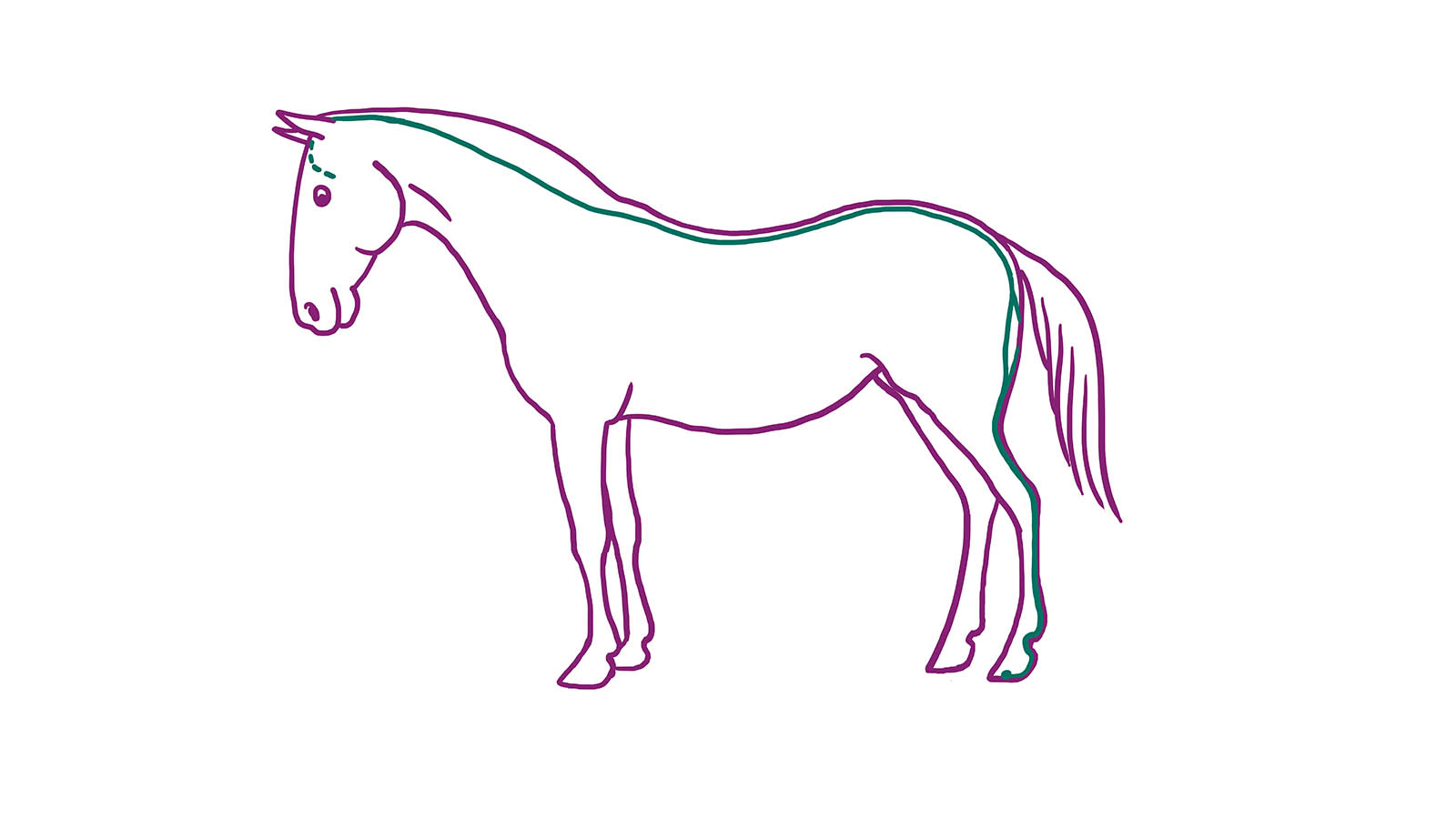
Superficial dorsal line
The superficial dorsal line has its origin in the hind limb phalanx and under the back hooves while the deep hamstring’s attachment, follow the tarsus, hip, on the back and attach behind the jaw.
Superficial ventral line
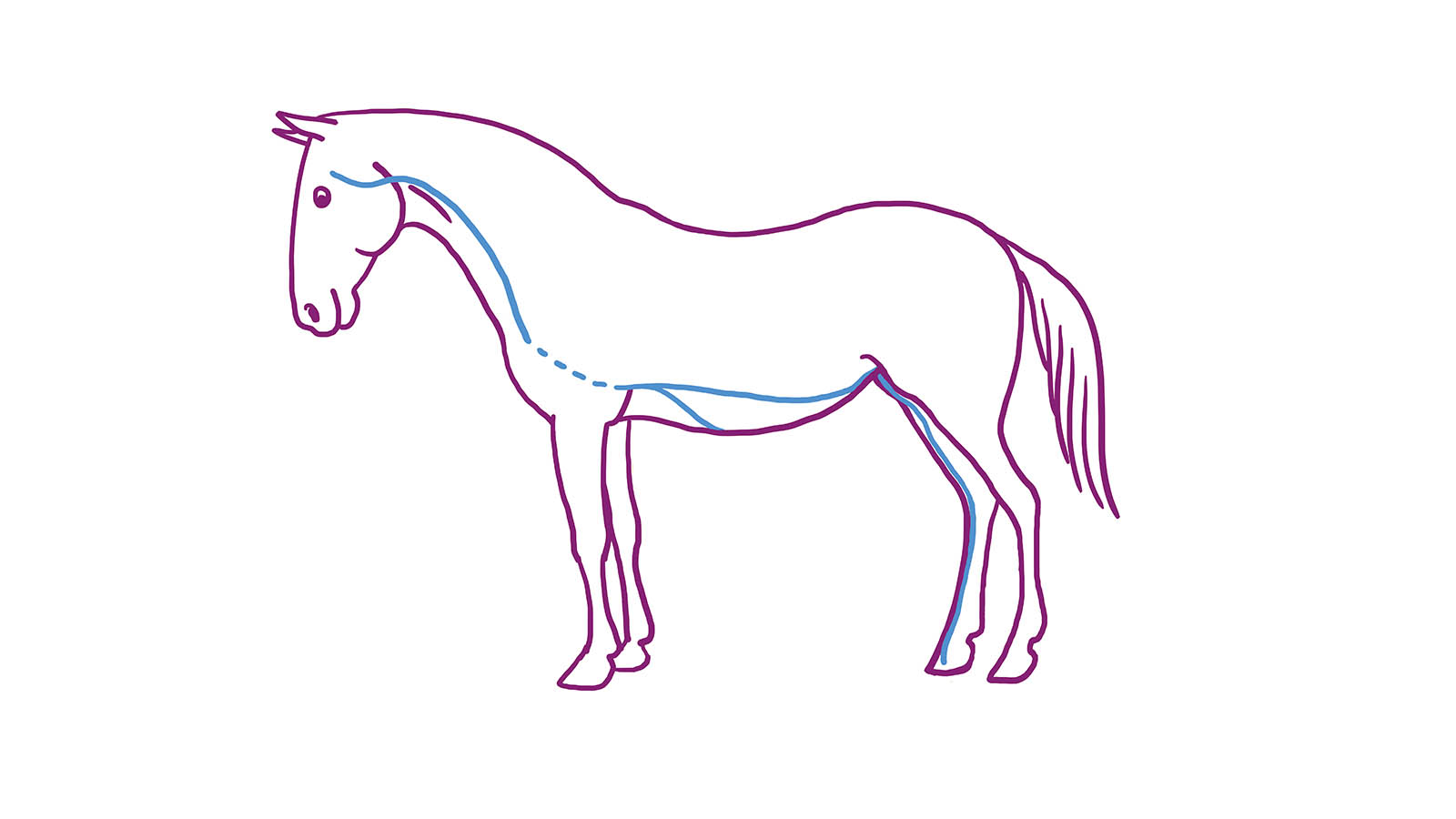
Superficial ventral line has its origin in the front of the hind limb in the outer phalanx the (coffin bone) goes up, past hocks front, to the knee, through the pelvis to the abdomen, the breast bone of it and on to the m. extensor and up to the jaw.
Lateral line
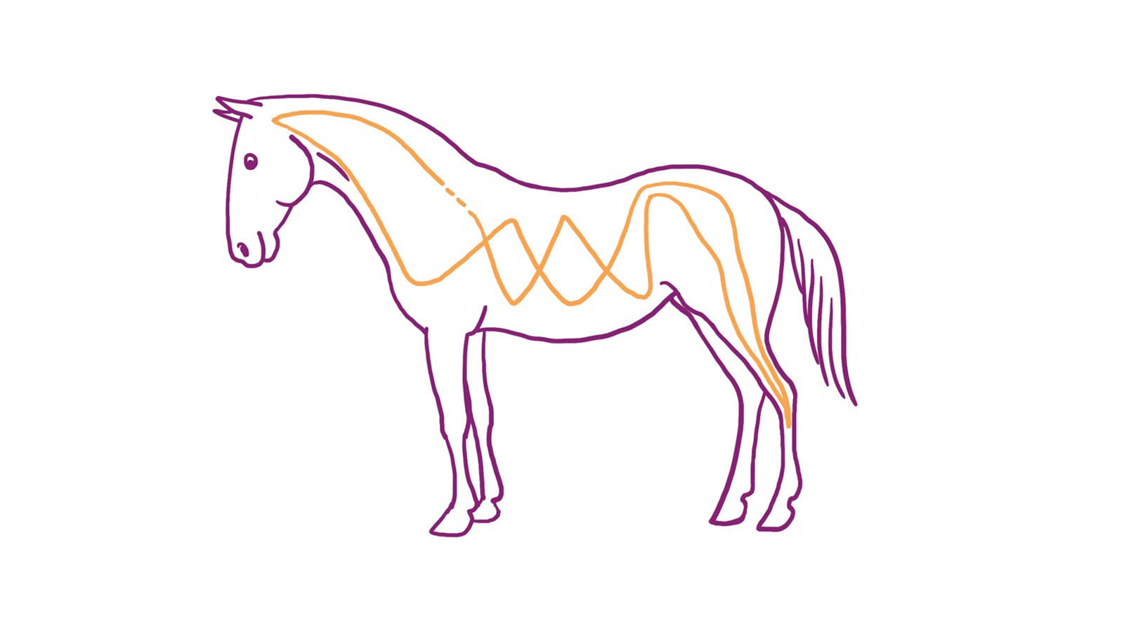
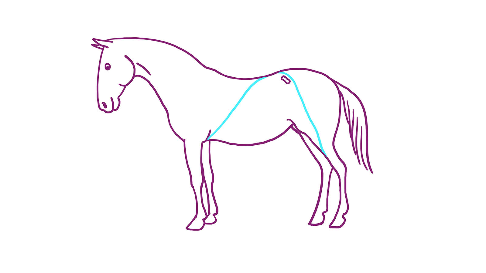
The lateral line originates in the hind limb of the coffin bone in the same area as the superficial ventral line. By the hock these two lines split up, and the lateral line goes up by the tensor fascia lata (outside of the thigh) to the tuber coxae (cross state) by m. Gluteus superficalis.
This is where this line is different from the others by splitting itself in a more superficial and deep line, although it still extends in the superficial layers. Through the core the lateral line extends in a cross pattern on the inside the shoulder blades. This line merges with m. Cutaneus trunci, the muscle that “shakes away the flies” on the horse’s sides.
The line then runs through m. Brachiocephalicus (lower neck muscles) up to the temple.
Spiral line
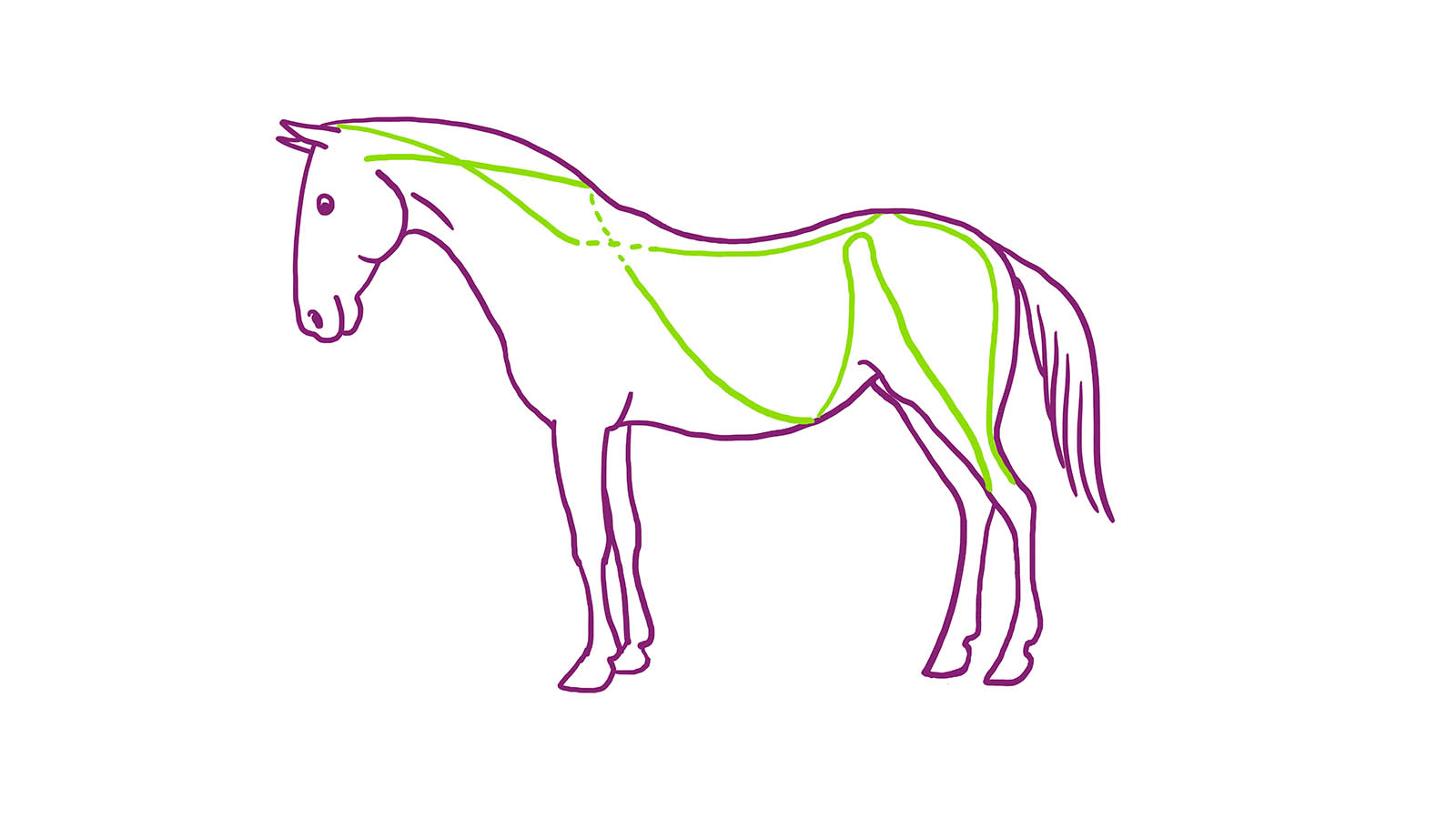
The spiral line has its origin cranially, at the temples and jaw joint and moves along the neck and thoracic spine, to m. rhomboids front and back part (withers / base of the neck) to move back to the tuber coxae, the line is aimed at the hock and continues through the biceps femoris, the sacrum (the cross) where the lines on either side cross over the back and then follows the superficial dorsal line up to the temple.
Functional line
Originated in the humerus (upper arm) crosses the chest over to the opposite side and down the outside of the thigh and knee where it merges with the patellar tendon (tendon in kneecap).
Front limb lines
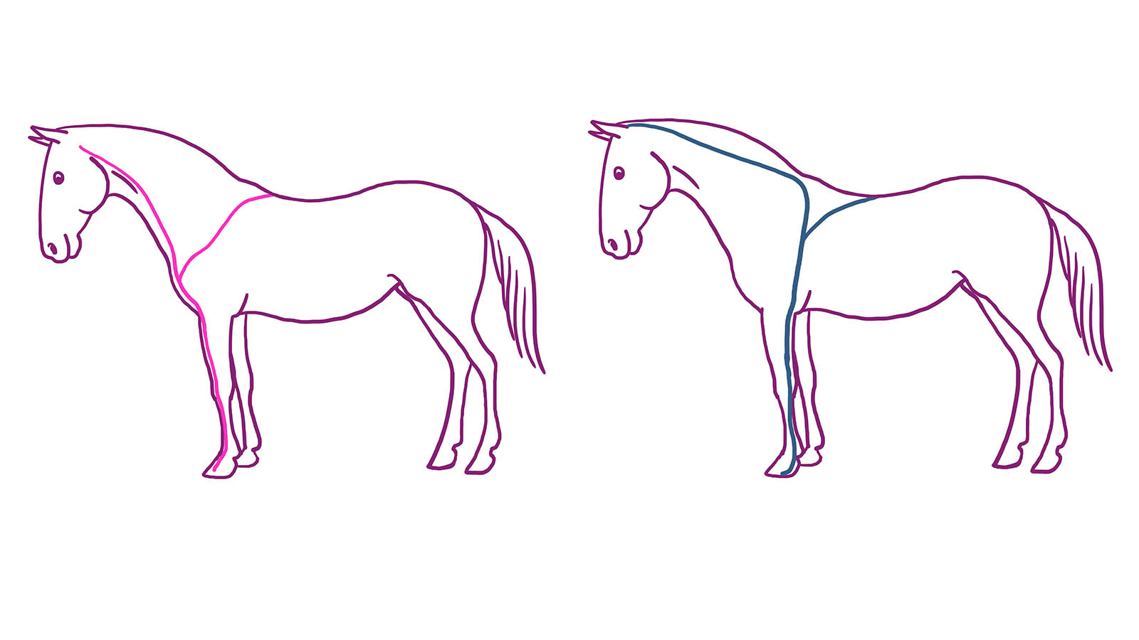
Two lines, one foreleg-stretching line.
Originating from the deep fascia at the skull, even from the deep cervical (neck) the fascia’s fascia pectoralis (chest muscles) from the deep fascia chest fascia and fascia auxiliaries (armpit)
Line two foreleg line’s cranial part.
From the skull via m. Brachiocephalicus to the shoulder blade’s m. Supraspinatus (frontal long superficial muscle on the outside of the scapula)
Onwards, up to m. Trapezius and m. Rhomboids
Discussion
The study reveals that full-body kinetic lines are present in the horse. The lines turned out to be very similar to those found in humans as described by Myers although clear differences were found.
The difference is due to the anatomical differences in the two and four-legged creatures.

